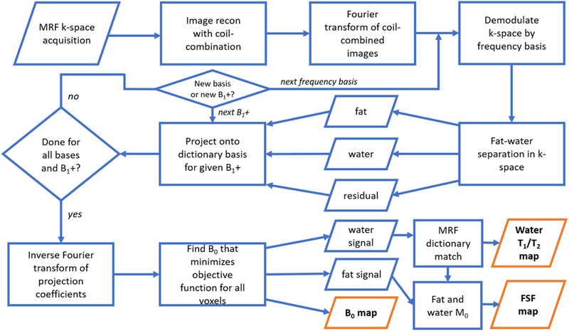
Fig. 1.

A flowchart of the implementation for the proposed MR fingerprinting (MRF) fat-water separation technique. A fixed-TR, linear swept TE is used for the MRF acquisition. Following gridding and coil-combination, the MRF stack is transformed back into k-space and demodulated as described in the Theory section. Fat-water-residual separation is performed in k-space for each demodulation frequency and each discretized B1+. The water, residual, and deblurred fat components are projected onto an approximate basis of a dictionary that includes T1, T2, and off-resonance effects. The coefficients of this projection are transformed to the image domain and smoothed (not shown), and B0 at each voxel is fitted. The B0 estimate is then used to appropriately combine the water and fat coefficients to yield fat signal fraction (FSF), and water T1 and T2 maps.
