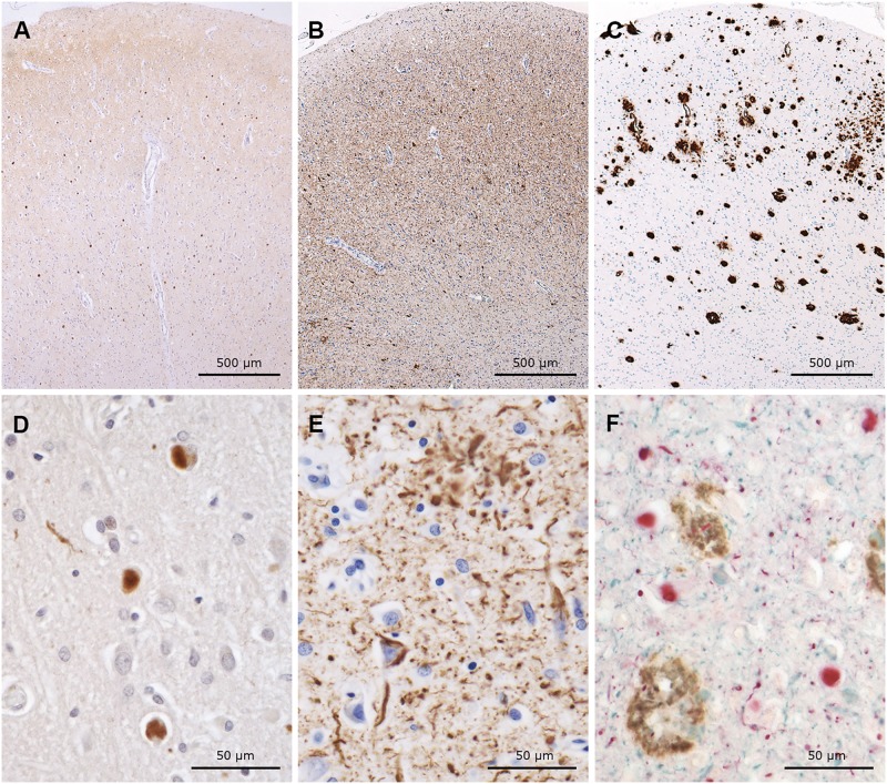
FIGURE 1.
Immunohistochemical preparations of serial sections from cingulate gyrus, labeled with antibodies to α-synuclein (A, D, brown), tau (B, E, brown), and Aβ (C, brown) as well as a triple immunohistochemical preparation using all 3 (F, α-synuclein = red, tau = blue, Aβ = brown). Note the numerous Lewy bodies (A, D, F), severe tau pathology (B, E, F), and Aβ pathology (C, F). (F) Photomicrograph shows the coexistence of all 3 abnormal proteins in the same section.

