
FIGURE 2.
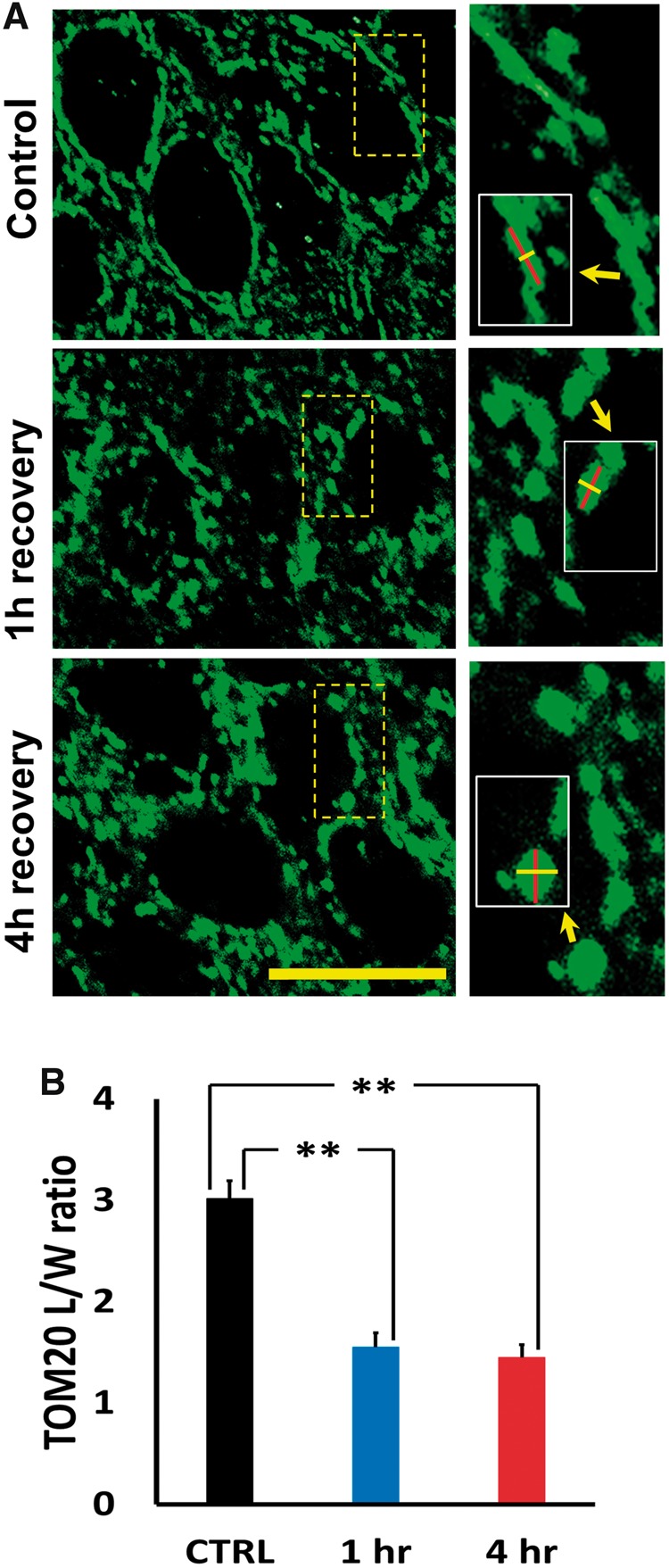
Ischemia disrupts mitochondrial morphology in CA1 pyramidal neurons. Rats were subjected to TGI followed by 1- or 4-hour recovery. Hippocampal slices were immunostained using antibody against the mitochondrial outer membrane marker, TOM20, and examined under confocal microscopy (1000×). (A) Representative images of TOM20-labeled CA1 mitochondria. Note the generally elongated mitochondria seen in perinuclear regions of control, in contrast to the abundant fragmented and swollen mitochondria 1 or 4 hours after ischemia. Small images of regions indicated show high magnifications of representative perinuclear mitochondria; inserts in small images indicate markings for length and width measurements of representative mitochondria (Bar = 10 µm). (B) Ischemia impacts mitochondrial morphology. Lengths and widths of individual mitochondria were measured blindly (using ImageJ software) and length/width (L/W) ratios calculated to quantify morphological change (lower L/W ratios indicates rounding of mitochondria). The L/W ratio for all mitochondria measured in neurons from a single animal was averaged to produce a single, mean L/W ratio for that animal. Note the significant decrease in L/W ratio after ischemia. Bars represent mean L/W ratio ± SEM from 4 to 6 independent animals each condition (comprising 125 mitochondria from 38 cells, control; ≥250 mitochondria, from ≥60 cells per group after TGI; ** indicates p < 0.01 by one-way ANOVA with Tukey post hoc).