
FIGURE 3.
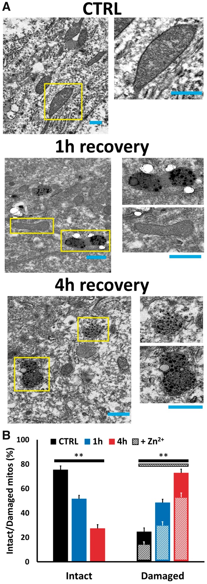
Global ischemia induces progressive Zn2+ accumulation and injury in CA1 pyramidal neuronal mitochondria. Rats were subjected to TGI followed by 1- or 4-hour recovery. To assess mitochondrial damage and Zn2+ accumulation in CA1 pyramidal neurons, Timm’s-labeled hippocampal slices were examined under TEM. (A) Representative electron micrographs. Representative TEM images (4000×) of Timm’s-labeled slices from a control rat (top), or rats subjected to ischemia followed by 1 hour (middle), or 4 hours (bottom) recovery (Bar = 0.5 µm; rectangles show regions displayed at greater magnification, right; Bar = 0.5 µm). Note the intact structure and absence of Timm’s precipitate in most mitochondria in control, the significant numbers of mitochondria showing early damage (with rounding) and distinct presence of Timm’s precipitate with 1-hour recovery, with greater numbers of mitochondria displaying Zn2+ accumulation and extensive injury after 4-hour recovery. (B) Quantitative evaluation. To assess mitochondrial Zn2+ accumulation and damage, all evident mitochondria in images from CA1 pyramidal neurons were rated by an observer blinded to the experimental condition. Bars display percentage of mitochondria appearing intact or showing evidence of damage (assessed as described), and hatchmarks indicate the presence of evident Zn2+ deposits in the damaged mitochondria (Zn2+ deposits were only present in <1% of intact appearing mitochondria in all conditions). Note the marked increase in numbers of damaged appearing mitochondria after ischemia, the marked increase in their numbers with increased recovery duration (from 1 to 4 hours), and the parallel increase in numbers containing distinct Zn2+ deposits, present in the majority of damaged mitochondria in each condition. Values represent mean ± SEM from 3 to 4 independent animals each condition (for each animal, ≥70 mitochondria from ≥10 sections were rated; ** indicates p < 0.01 by one-way ANOVA with Tukey post hoc).