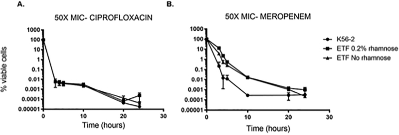
Figure 2.
Persister cells formation of the CGetf mutant strain. The percentage of viable cells (y-axis) was calculated after treatment with 100 μg/mL ciprofloxacin (A) or 800 μg/mL meropenem (B) of exponentially growing cells of, wild type K56–2 (black circles) and CGetf (ETF) in the presence (black squares) or absence (black triangles) of 0.2% rhamnose. X-axis indicates time in hours. Time 0 indicates the initial % of viable cells before treatment. An aliquot of each culture was taken at 3, 4, 5, 20 and 24 hours’ post addition of antibiotic, washed with LB and plated for colony counting. The graphs include standard deviation (SD) of three biological replicates.

