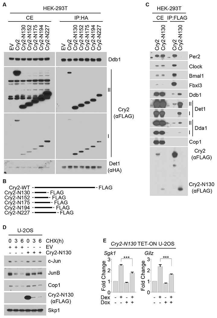
Figure 5. The N-terminus of Cry2 interacts with Det1 to stabilize Cop1’s substrates and inhibit GR activity.

(A) HA-tagged Det1 was co-expressed in HEK-293T cells with either FLAG-tagged wild-type Cry2 or FLAG-tagged Cry2 truncation mutants followed by immunoprecipitation from cell extracts with an anti-HA resin. Cells extracts (CE) and immunoprecipitates (IP) were analyzed by immunoblotting. Roman numbers indicate different exposure times: I, short exposure; II, long exposure.
(B) Schematic representation of wild-type Cry2 and Cry2 truncation mutants.
(C) FLAG-tagged Cry2 or FLAG-tagged Cry2-N130 truncation mutant were expressed in HEK-293T cells and immunoprecipitated from cell extracts with an anti-FLAG resin. CEs and immunoprecipitates were then analyzed by immunoblotting.
(D) U-2OS cells were transfected with either an empty vector (EV) or with a vector expressing Cry2-N130. 18 hours after transfection, cells were treated with CHX for the indicated times and cell extracts were immunoblotted.
(E) qPCR analysis of cDNAs prepared from U-2OS cells infected with lentiviruses expressing Cry2-N130 under the control of a doxycycline-inducible promoter and treated with dexamethasone where indicated. See also Figures S3, S4 and S5.
