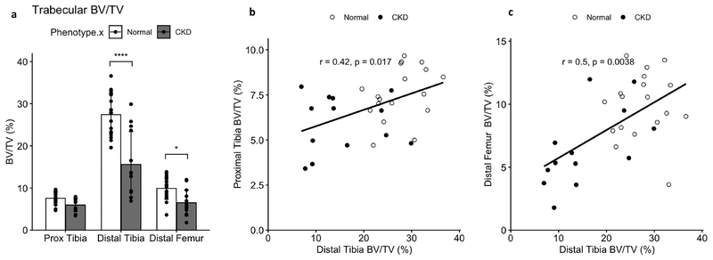
Figure 7 -.
Trabecular bone volume at the proximal tibia, distal tibia, and distal femur sites. CKD animals had significantly reduced BV/TV (a) compared to NL at both distal locations. Spearman correlations confirmed significant relationships between the distal tibia BV/TV and BV/TV at both the proximal tibia (b) and distal femur (c), supporting the usefulness of the distal tibia for monitoring BV/TV changes in CKD over time. Bar plots show mean +/− SD. *p <= 0.05; **p <= 0.01; ***p <= 0.001; ****p <= 0.0001

