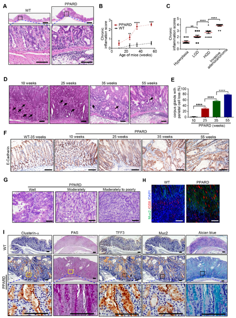
Figure 2. Villin promoter–driven PPARD overexpression induced chronic inflammation and premalignant changes in mice.

(A) Representative H&E-stained images of chronic inflammation induced in gastric corpus of PPARD mice in contrast to their WT littermates at 35 weeks.
(B, C) Gastric corpus inflammation scores of PPARD and WT mice by age (B) and morphology (C).
(D, E) Representative H&E images of gastric corpus from PPARD mice showing parietal cells indicated by black arrows (D) and their quantitation, analyzed for 50 glands per mouse (E). n=15-20 per group.
(F) Representative E-cadherin IHC microphotographs in gastric corpus of PPARD mice and WT mice at the indicated ages.
(G) Representative images of PPARD–induced gastric adenocarcinoma differentiation in PPARD mice at 55 weeks.
(H, I) Representative microphotographs of GSII and Muc2 immunofluorescence (H) and clusterin-α, TFF3, and Muc2 IHC and of PAS and alcian blue staining (I) in the gastric corpus of PPARD mice and WT littermates at 35 weeks.
Scale bars, 1 mm (A [top]) and 100 μm (A [bottom], D, F, G-I).
