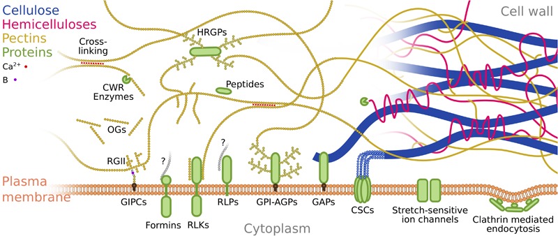
FIGURE 2.

Putative mechanosensing structures at the cell wall and plasma membrane. Schematic representation of the various structures potentially involved in plant mechanosensing, and their interactions. For clarity and ease of representation we focus on some of the interactions between pectins, proteins and plasma membrane (and focus less on cellulose and hemicelluloses). Cellulose is represented in blue, hemicelluloses in magenta, pectins in yellow, proteins in green and plasma membrane in orange. Red dots represent calcium ions involved in pectin cross linking. The purple dot represents boron involved in RGII-GIPC cross-linking. Gray polysaccharides labeled with a question mark in the case of Formins and RLPs accounts for interaction of these proteins with the cell wall with the nature of the polysaccharide or molecule involved remaining unknown. Ca2+, Calcium; B, Boron; CWR, Cell Wall Remodeling; HRGP, Hydroxyproline-rich Glycoproteins; OGs, OligoGalacturonans; RGII, RhamnoGalacturonanII; GIPCs, Glycosyl Inositol Phospho Ceramides; RLKs, Receptor-Like Kinases; RLPs, Receptor-Like Proteins; GPI-AGPs, GlycosylPhosphatidylInositol anchored ArabinoGalactan Proteins; GAPs, GlycosylPhosphatidylInositol Anchored Proteins; CSCs, Cellulose Synthase Complexes.
