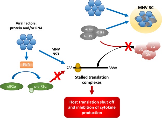
FIG 7.
Model. During MNV infection, viral factors such as proteins and/or RNA (blue hexagon) phosphorylate eIF2α (green oval) via PKR (orange rectangle), as well as stalling translation initiation by the MNV NS3 protein. However, this translational arrest is uncoupled from the PKR–p-eIF2α axis. These stalled preinitiation complexes typically aggregate with G3BP1 (gray oval) and form SGs (red cloud). However, MNV viral factors sequester G3BP1 to the MNV RC (yellow circle) to promote replication. This allows the inhibition of cap-dependent host cell translation, as well as inhibiting the formation of SGs.

