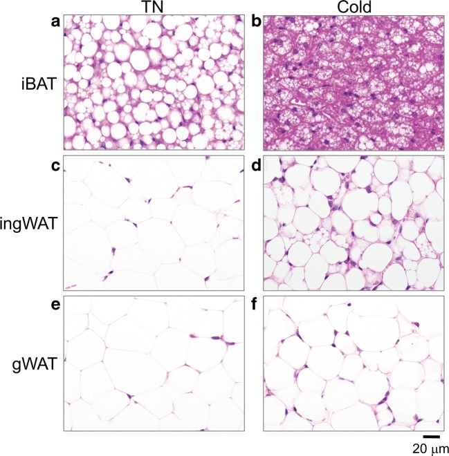
Fig. 2.
a–f Representative bright field images of H+E stained sections of interscapular brown adipose tissue (iBAT), inguinal white adipose tissue (ingWAT) and gonadal white adipose tissue (gWAT) from mice acclimated to cold (4 °C) compared to thermoneutrality (30 °C, TN). All images were taken at × 600 magnification

