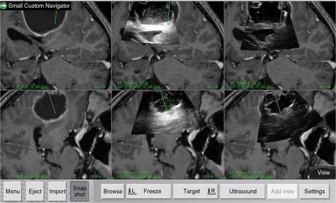
Fig. 2.
Display from the navigation system, showing two perpendicular image slices extracted from the MRI or 3D ultrasound volume according to the orientation of the navigated tool. The preoperative MR T1-weighted contrast image is shown to the left. The middle column shows the ultrasound images acquired with Ringer’s solution in the resection cavity, and in the right column, the ultrasound obtained with ACF-3 is shown overlaid the MR images

