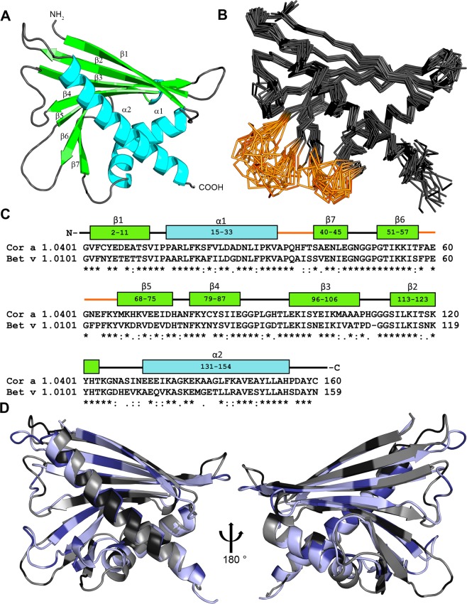
Figure 1.
Solution structure of Cor a 1.0401. (A) Cartoon representation of the average of the 20 lowest energy solution structures of Cor a 1.0401 (PDB: 6GQ9) (α-helices, turquoise; β-strands, green; loop-regions, grey). (B) Backbone overlay of the 20 lowest energy solution structures of Cor a 1.0401, with a backbone rmsd value of 0.93 Å and overall rmsd of 1.26 Å. The loop regions Ala35 - Thr40 (between strand β7 and the two short α-helices) and Thr58 - Met68 (between strand β5 and β6) which did not show resonances are highlighted in brown. (C) Amino acid sequence alignment of Cor a 1.0401 and Bet v 1.0101. The α-helices and β strands of Cor a 1.0401 are shown in blue and green, respectively, the two loop regions are highlighted in brown. (D) Overlay of the structures of Cor a 1.0401 (grey) and Bet v 1.0101 (light blue) (PDB: 6R3C). Black and dark blue indicate sequence differences.

