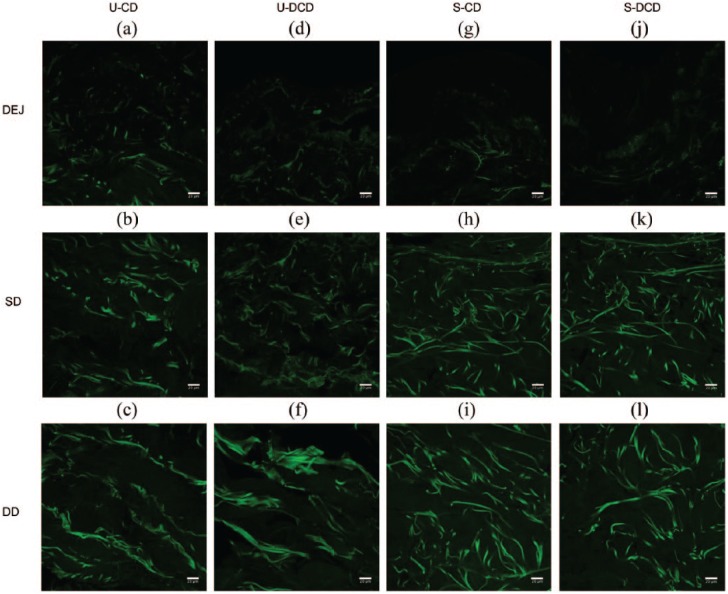
Figure 4.
High-resolution 2D multiphoton images of collagen. High-resolution large-area SHG images (512 × 512 pixels, 8-bit) were obtained by MPM from unscarred and scarred dermis, both pre- and post-decellularisation. SHG images were extracted from the channel corresponding to wavelengths in the range of 398–409 nm, with an excitation wavelength of 810 nm and operating at 15% of maximum laser power. Images were obtained at differing levels across the longitudinal length of the sample. Images presented are from a single patient for ease of illustration. The 2D configuration of collagen fibers in unscarred (a-f) and scarred (g-l) dermis is visible by the intense green SHG signal. Few collagen fibers were visible at the DEJ of each specimen (a, d, g, j). Scale bars: 20 μm.
U-CD: unscarred cellular dermis; U-DCD: unscarred decellularised dermis; S-CD: scarred cellular dermis; S-DCD: scarred decellularised dermis; DEJ: dermal–epidermal junction; SD: superficial dermis; DD: deep dermis; MPM: multiphoton microscopy; SHG: second harmonic generation.

