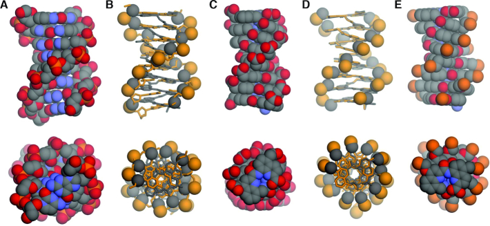
Figure 6.
Side views and top views shown at the same scale of: (A) crystal structure of 8-bp B-DNA duplex d(CGCTAGCG)2 (PDB ID: 250D) (70); (B) molecular overlay of B-DNA (ochre) and DNA mimic 4 (gray); (C) crystal structure of DNA mimic 4; (D) molecular overlay of DNA mimic 4 (gray) and a molecular model of (mQPhoQ5Pho)8 (ochre) and (E) a molecular model of (mQPhoQ5Pho)8. In (B) and (D), foldamers or DNA are shown in stick representations, except the side chain carboxylate carbon atoms, and the phosphonate and phosphate phosphorus atoms that are shown as spheres. Two molecules of (mQPhoQ5Pho)4 as observed in the crystal (33) have been stacked to produce the model of (mQPhoQ5Pho)8. Hydrogen atoms have been removed for clarity. In (E), ethyl ester functions at the side chains, tert-butyl-carbamate and benzyl ester functions at the N terminus and C terminus, respectively, have been removed for clarity.

