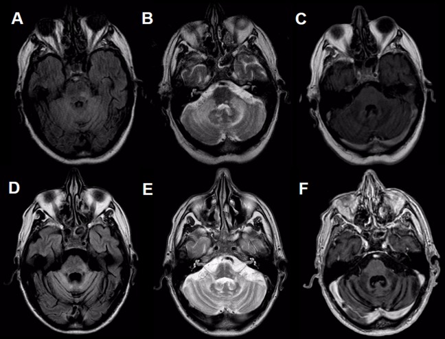
Figure 1.
Magnetic resonance imaging of the brain. Hyperintensity on FLAIR (A, D), y T2 (B, E), and hypointensity on T1 (C, F) in both cerebellar hemispheres and peduncles with extension to the pons, predominantly at the level of the left posterolateral sector, without contrast enhancement. Figures A-C correspond to 6 months after initial symptoms, and Figures D-F, after 17 months. FLAIR indicates fluid-attenuated inversion recovery.

