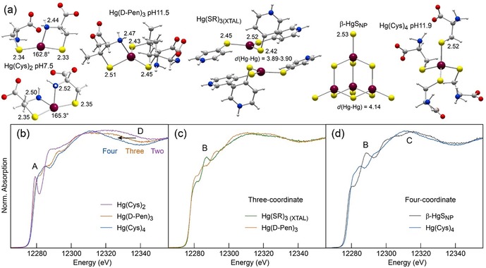
Figure 4.

Sensitivity of L3‐edge HR‐XANES to the bonding environment of Hg. (a) Structure of the Hg complexes. (b–d) Spectra of two‐, three, and four‐coordinate Hg‐thiolate and Hg‐sulfide references. The Hg(Cys)2 complex was prepared at a Cys:Hg molar ratio of 2 and pH 7.5, the Hg(d‐Pen)3 complex at d‐Pen:Hg=10.0 and pH 11.5, and Hg(Cys)4 at Cys:Hg=10.0 and pH 11.9. Their structure was optimized geometrically (MP2‐RI/def2‐TZVP‐ecp67), and the Hg(SR)3 complex and β‐HgSNP model are X‐ray crystal structures. Peak B is diagnostic of Hg–Hg pairs. Bond lengths are in angstroms. Dark red, Hg; yellow, S; blue, N; red, O; gray, C; light gray, H.
