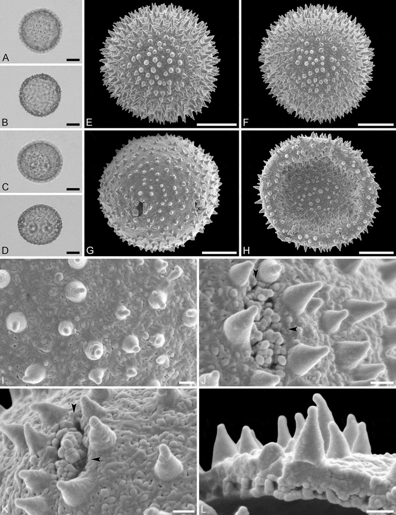
Figure 6.

Light microscopy (A–D) and scanning electron microscopy (E–L) micrographs of Hyaenanche globosa (from South Africa, coll. unknown, s.n. [WAG: E, F, H, J, L]; from Namibia, coll. Hall, 3912 [PRE: A–D, G, I, K]). A. Polar view, high focus. B. Polar view, optical cross-section. C. Equatorial view, high focus. D. Equatorial view, optical cross-section. E. Polar view. F. Polar view. G. Equatorial view. H. Infolded grain. I. Close-up of interapertural area. J. Close-up of aperture, showing membrane (arrows). K. Close-up of aperture, showing membrane (arrows). L. Close-up showing section through pollen wall. Scale bars – 10 µm (A–H), 1 µm (I–L).
