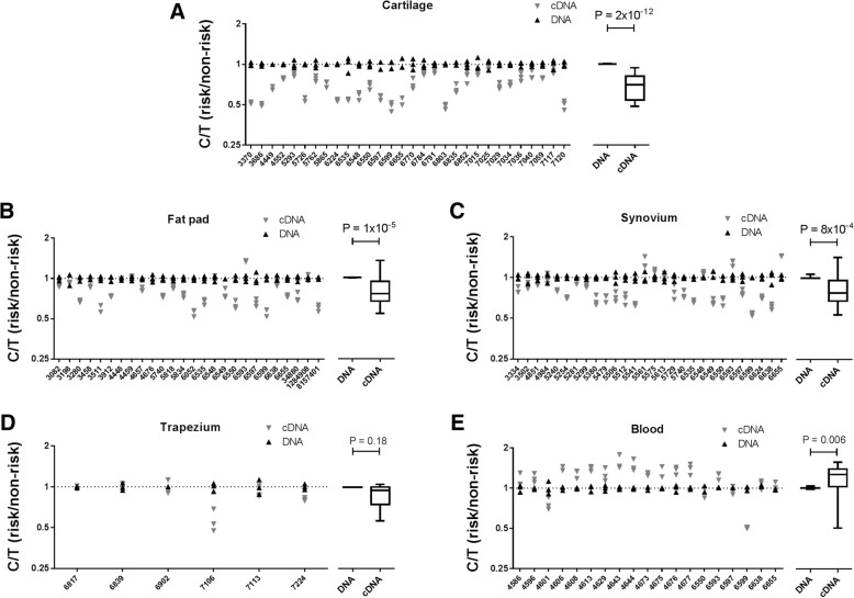
Fig. 2.
Allelic expression imbalance (AEI) analysis of MGP. AEI analysis of rs4236 was carried out in OA patient cartilage (n = 30) (a), infrapatellar fat pad (n = 26) (b), synovium (n = 28) (c), trapezium (n = 6) (d) and blood (n = 19) (e). The y-axis for each plot shows the risk/non-risk (C/T) allelic ratios, with a ratio < 1 indicating decreased expression and a ratio > 1 indicating increased expression of the C allele. Three technical repeats were performed for each patient’s DNA and cDNA. The right panels show the mean values for DNA and cDNA from all patients combined, with results represented by box-and-whisker plots, in which the lines within the box represent the median, the box represents the 25th to 75th percentiles, and the whiskers represent the minimum and maximum values. P values were calculated using a Mann-Whitney 2-tailed exact test. Individual patients are designated by their anonymised identification (ID) numbers

