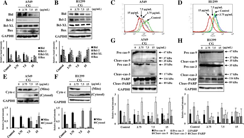
Fig. 6.
Effects of CG on MMP and intrinsic apoptosis pathway-related factors in A549 and NCI-H1299 cells. Protein expression of BID, Bcl-2, Bcl-xL, Bax, and GAPDH in A549 (a) and NCI-H1299 (b) cells, as determined by western blotting. Cells were treated with various doses of CG for 48 h and compared with untreated cells. Histogram profiles of JC-1 aggregates (FL-2, orange) detected by flow cytometry of A549 (c) and NCI-H1299 (d) cells. Western blotting of cytochrome c protein in the mitochondria and the cytosol, and GAPDH in A549 (e) and NCI-H1299 (f) cells. Protein expression of the intrinsic pathway factors, caspase-9, caspase-3, PARP, and GAPDH in A549 (G) and NCI-H1299 (H) cells, as determined by western blotting. The data are presented as the mean ± SEM (n = 3). The data were analyzed using one-way ANOVA with Tukey’s HSD test. *, p < 0.05 and **, p < 0.005. Cyto c, cytochrome c; Mito, mitochondria; Pro cas-9, pro-caspase-9; Cleav-cas-9, cleaved caspase-9; Pro cas-3, pro-caspase-3; Cleav-cas-3, cleaved caspase-3; Cleav-PARP, cleaved PARP

