Fig. 1.
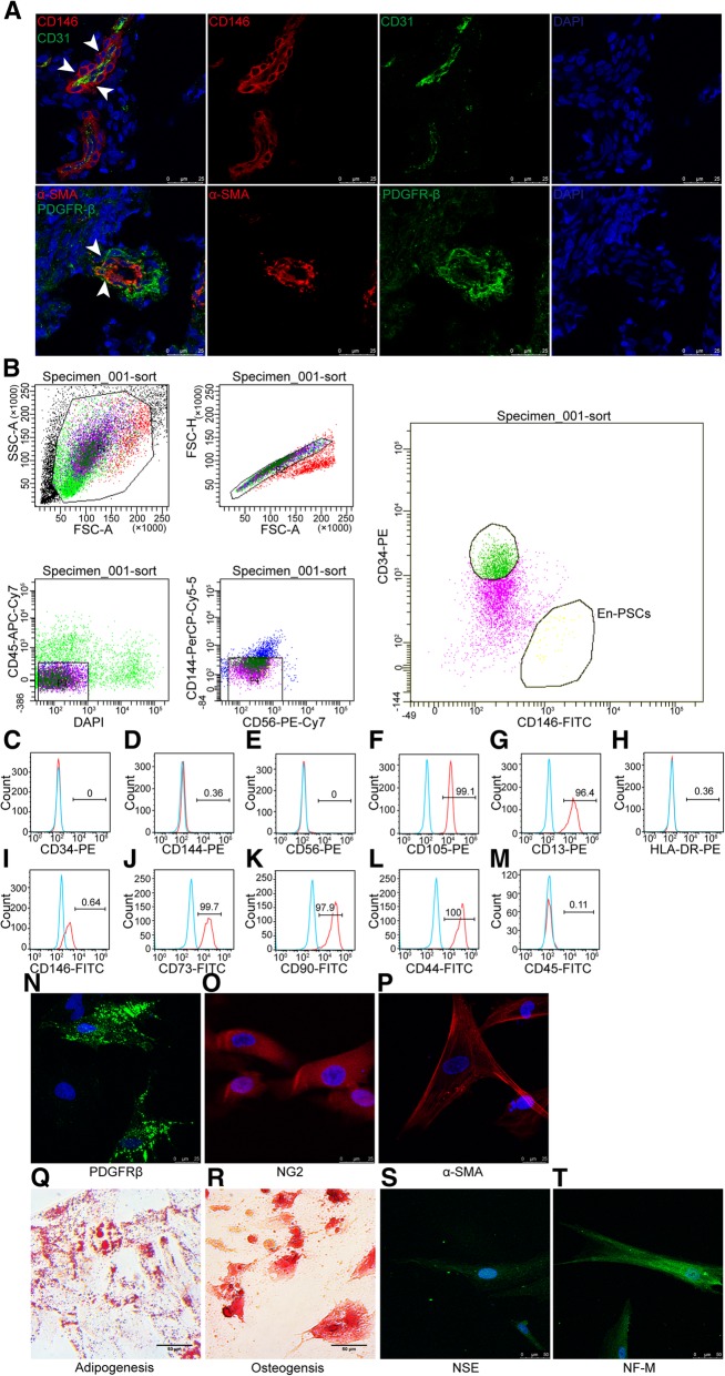
Phenotype and multidirectional differentiation potential of En-PSCs. a Immunofluorescence staining in human endometrium displayed CD146, CD31, PDGFRβ, and α-SMA. The arrow showed En-PSCs. Scale bars, 25 μm. b En-PSCs (CD146+CD34-CD45-CD56-CD144-) were sorted by flow cytometry. c–m Flow cytometry analysis of immune-markers in En-PSCs. n–p En-PSCs expressed PDGFRβ, NG2, and α-SMA. Scale bars, 25 μm. q Intracellular lipid droplet induced from En-PSCs were detected by Oil red O staining. Scale bars, 50 μm. r Calcified nodule stained with Alizarin red S indicated that En-PSCs differentiated to osteogenic lineage. Scale bars, 50 μm. Neural-like differentiation was confirmed by immunofluorescence staining with anti-NSE antibody (s) and anti-NF-M antibody (t). Scale bars, 50 μm

