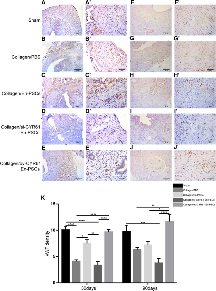
Fig. 5.
Immunohistochemical staining of vWF in the newly regenerated uteri after transplantation of collagen/transfected En-PSCs. Detection of vWF expression for blood vessels in the regenerated uteri at days 30 days (A–E) and 90 days (F–I) in sham group (A, F), collagen/PBS group (B, G), collagen/si-CYR61 En-PSCs group (C, H), collagen/vector En-PSCs group (D, I), and collagen/ov-CYR61 En-PSCs group (E, J). Arrowheads indicate repair site. Scale bar, 500 μm. Local magnification of figures (A–J) were showed in the pictures (A′–J′). Scale bar, 50 μm. K The statistical analysis of the capillary vessels number from at least three randomly selected fields under a magnification of 400. Each experiment was repeated four times. Data were presented as mean ± SEM. *P < 0.05, **P < 0.01, ***P < 0.001, and ****P < 0.0001

