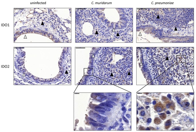
Figure 4.
Detection of IDO1-2 protein expressions in Chlamydia infected and uninfected BALB/c mouse lungs. IDO1 protein and IDO2 protein expressions detected by immunohistochemistry in C. muridarum infected, C. pneumoniae infected (7 days post infection) and uninfected control lung tissues. The IDO positive epithelial cells are shown by gray triangles, the IDO positive macrophages are shown by black triangles. Bars: 50 μm. The characteristic IDO stainings of epithelial cells and macrophages are shown in brackets. Bars: 5 μm.

