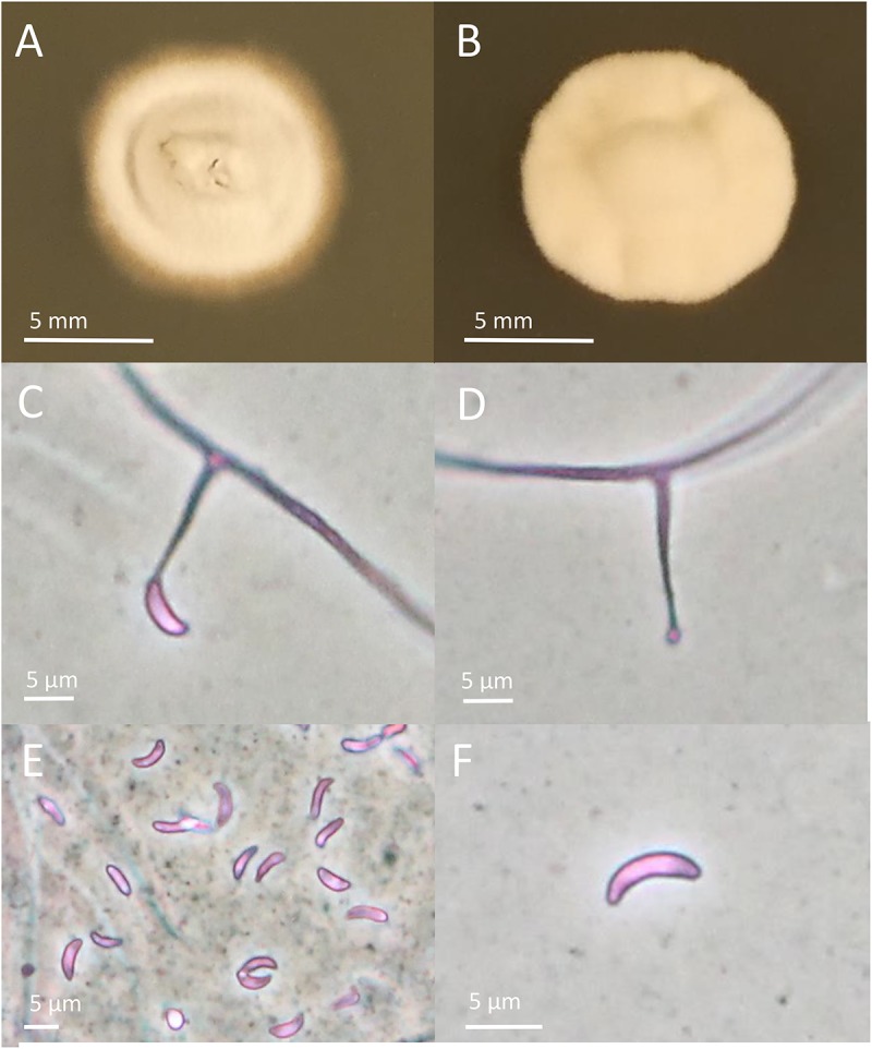
FIGURE 1.

Colony and conidial morphology of Epichloë isolates from Schedonorus arundinaceus var. glaucescens. (A,B) Different colony morphologies of FaTG-5 isolates. (C,D) Conidiophores from the two isolates. (E,F) Conidia from the two isolates. Pictures of colonies were taken after growing on PDA for 4 weeks.
