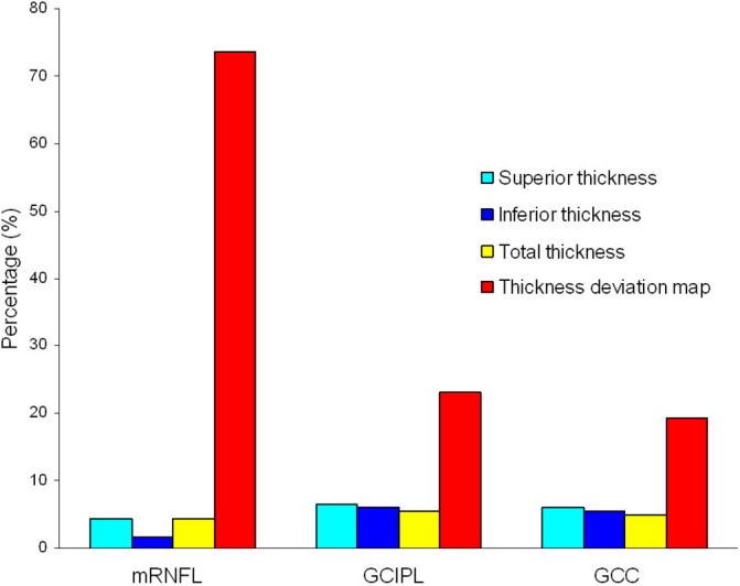
Figure 1.

Percentages of eyes classified as abnormal according to the four optical coherence tomography parameters (superior thickness, inferior thickness, total thickness and cluster in thickness deviation map) for the macular retinal nerve fibre layer (mRNFL), ganglion cell-inner plexiform layer (GCIPL) and ganglion cell complex (GCC).
