Figure 2.
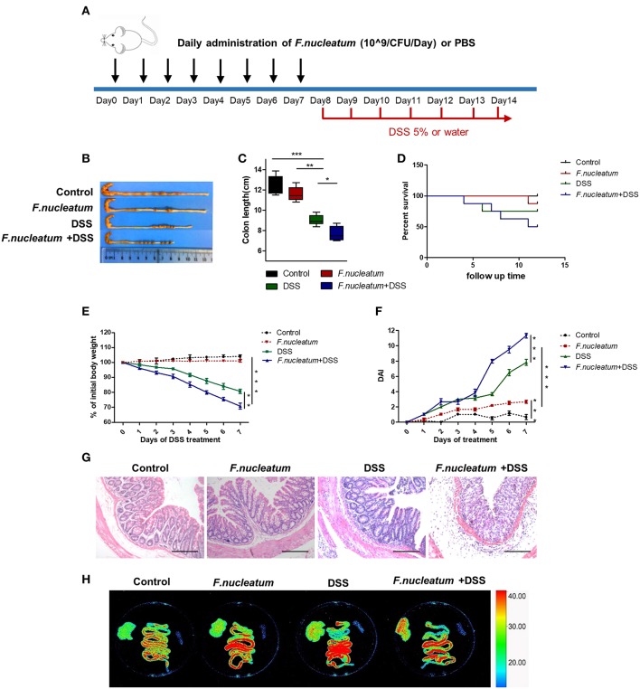
F. nucleatum increased the susceptibility of mice to DSS-induced experimental colitis. BABL/C mice were subjected to a DSS-induced colitis induction protocol using 5% DSS in drinking water for 7 consecutive days. (A) F. nucleatum and DSS treatment schedule. (B,C) Macroscopic appearance and quantification of colon length was determined at day 7 after DSS induction of colitis, n = 6 mice per group. Representative images are shown. (D) Survival curves of mice in this system, n = 6 mice per group. (E) Body weights determined daily for all mice, n = 6 mice per group. (F) Daily DAI scores for all mice, n = 6 mice per group. (G) Serial sections of colon tissues were stained with H&E to investigate tissue damage. Representative images are shown. Scale bars, 50 μm. (H) Ex vivo images of the liver, spleen, and intestine 12 h after gavage of mice with ERFP-E.coli. Representative images are shown. Each experiment was performed at least three times. Data are presented as means ± SD. *P < 0.05; **P < 0.01; ***P < 0.001; Analysis of Variance (ANOVA) and Student's t-test (two-tailed).

