Figure 6.
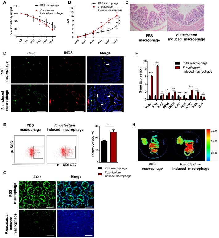
Adoptive transfer of F. nucleatum-induced macrophages aggravates colitis progression. F. nucleatum-treated BMDMs were collected and injected into the abdominal cavity of mice 48 h before initiation of DSS. (A) Body weight was measured daily, n = 6 mice per group. (B) DAI scores were recorded daily, n = 6 mice per group. (C) Mucosal tissues collected on day 5 were H&E stained, with representative images shown to assess colitis severity. Representative images are shown. Scale bars, 50 μm. (D) Mucosal tissues were stained by F4/80 (green) and iNOS (red) in macrophages; DAPI (blue) served as a nuclear stain. Representative images are shown. Scale bars, 50 μm. (E) F4/80 and CD16/32 staining of LP cells. Ratio of F4/80 and CD16/32 double positive cells were performed. (F) Expression of TNF-α, IFN -γ, IL-12, IL-10, MCP-1, Arg-1, iNOS, and ZO-1 was measured by qRT-PCR in colon tissues. (G) Mucosal tissues were stained with ZO-1 (green); DAPI (blue) served as a nuclear stain. Representative images are shown. Scale bars, 10 μm. (H) Ex vivo images of the liver, spleen and intestine 12 h after ERFP-E. coli gavage. Representative images are shown. Each experiment was performed at least three times. Data are presented as means ± SD. *P < 0.05; **P < 0.01; ***P < 0.001; Analysis of Variance (ANOVA) and Student's t-test (two-tailed).

