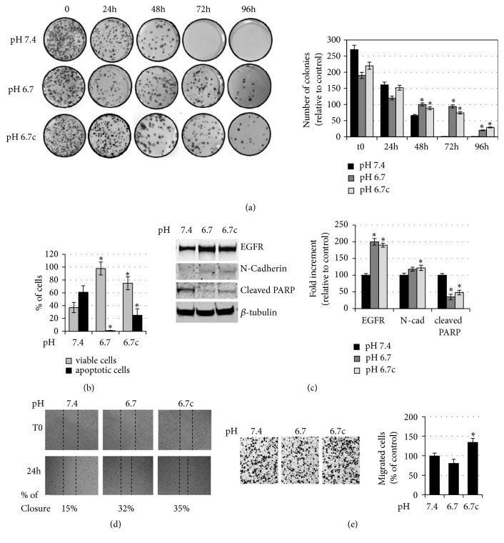
Figure 3.
Evaluation of the anoikis resistance of acidic melanoma cells grown in rocking conditions. (a) Representative images (left) of clonogenic efficacy in acidic and nonacidic A375M6 melanoma cells after exposure to 24-96 h of rocking condition. After treatment, the cells were seeded in 100 mm plates (5000 cells/dishes, in triplicate) and incubated for 12–14 days. Quantification (right) of cloning efficiency expressed as number of colonies. (b) Annexin V/PI flow cytometric analysis of acidic and nonacidic A375M6 melanoma cells after exposure to 48h of rocking condition; the vital cells were negative for both annexin V and PI staining; the total cell death was the sum of cells positive for annexin V, PI, or both stainings. (c) Representative images (left) of immunoblotting for EGFR, N-cadherin, cleaved PARP, and β-tubulin of acidic and nonacidic A375M6 melanoma cells and after exposure to 48h of rocking condition and (right) densitometry graph of protein expression. (d) Representative images (left) of the wound healing assay on acidic and nonacidic A375M6 melanoma cells after exposure to 48h of rocking condition. Images are taken immediately after scratching (t0) and 24 hours later (24 h). Percentage indicates wound closure compared to t0. (e) Representative images (left) of invasiveness of acidic and nonacidic A375M6 melanoma cells after exposure to 48h of rocking condition and quantitative analysis (right) of the number of cells that migrated through Matrigel. Each experiment was conducted in triplicate and data are expressed as mean ± SEM of at least three independent experiments. ∗ p<0.05.

