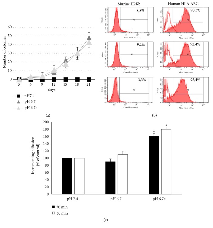
Figure 6.
In vivo evaluation of anoikis resistance of acidic melanoma cells. (a) Emerging colonies derived from blood sample obtained from mice (n=3) i.v. injected with acidic and nonacidic A375M6 melanoma cells; colonies with over 20 cells were counted. (b) Representative images of flow cytometer analysis of HLA-ABC or H2Kb antigen expression in pooled cell population that recovered from blood samples after 21 days. (c) Evaluation of adhesiveness of acidic and nonacidic A375M6 melanoma cells toward activated endothelial cells at 30 and 60 min through flow cytometer analysis of acidic and nonacidic CSFE-positive A375M6 melanoma cells and activated endothelial cells (CSFE-negative) cocultures. Images are representative of experiments performed in triplicate; data are expressed as mean ± SEM of at least three independent experiments. ∗ p<0.05.

