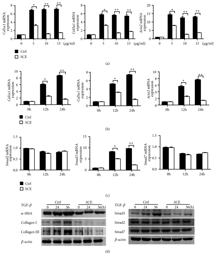
Figure 5.
SCE attenuates fibrotic responses in CFs by inhibiting TGF-β/Smad3 signaling activation. (a) RT-qPCR analysis of mRNA expression for genes of Col1a1, Col3a1, and Acta2 in cardiac fibroblasts (CFs) pretreated with different concentrations of SCE (5 μl/ml, 10 μl/ml, and 15 μl/ml) followed by TGF-β stimulation (20 ng/ml) for 24 h. (b) RT-qPCR analysis of mRNA expression for genes of Col1a1, Col3a1, and Acta2 in CFs pretreated with SCE (10 μl/ml) followed by TGF-β stimulation (20 ng/ml) for 12 h and 24 h. (c) RT-qPCR analysis of mRNA expression for genes of Smad2, Smad3, and Smad7 in CFs pretreated with SCE (10 μl/ml) followed by TGF-β stimulation (20 ng/ml) for 12 h and 24 h. (d) Immunoblot analysis of expression of Smad2, Smad3, and Smad7 in CFs pretreated with SCE (10 μl/ml) followed by TGF-β stimulation (20 ng/ml) for 24 h and 36 h. Data are from three independent experiments ((a)–(c), mean ± SEM) or are representative of three independent experiments with similar results (d). ∗P < 0.05 and ∗∗P < 0.01, Student's t-test.

