Figure 2.
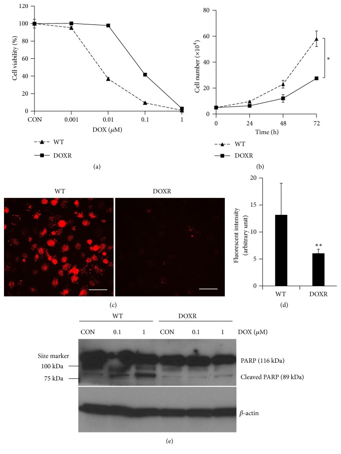
Characterization of DOXR cells. (a) DOX sensitivity of WT (IC50 = 8 nM) and DOXR (IC50 = 80 nM) cells. (b) Doubling time of WT and DOXR cells. ∗p < 0.05, two-tailed Student's t-test. (c) DOX efflux ability of WT and DOXR cells. White bar = 50 μm. (d) Red fluorescent intensity of WT and DOXR cells obtained by analyzing (c) using ImageJ software. ∗∗p < 0.01, two-tailed Student's t-test. (e) PARP and cleaved PARP protein expression of DOX treated WT and DOXR cells. DOX treated for 48 h. Control (CON) groups were treated in growth medium with 0.01% DMSO (v/v) as vehicle. β-Actin was used as endogenous loading control.

