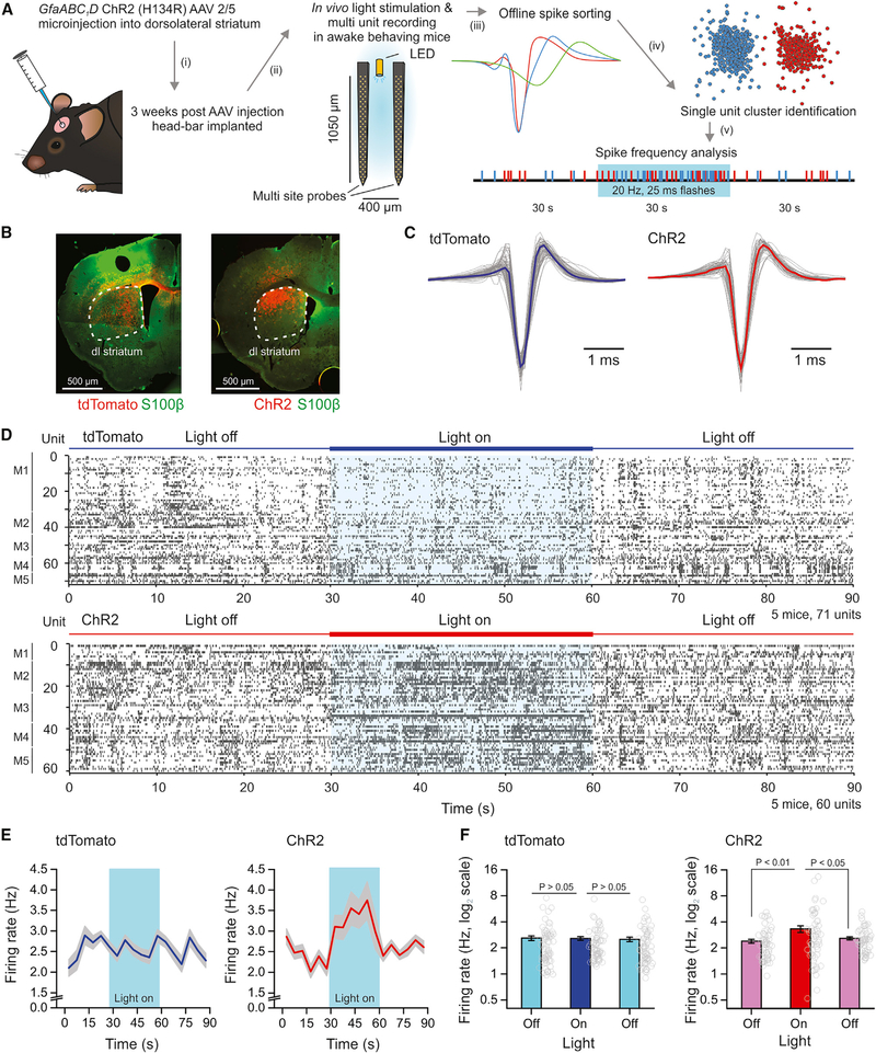
Figure 5. In Vivo Activation of ChR2 in Astrocytes Enhances MSN AP Firing Measured with Silicon Probes in Awake Behaving Mice.
(A) Schematic depicting experimental workflow using multi-site silicon probes in vivo. Mice were injected with AAVs to express tdTomato or ChR2 within astrocytes. Light flashes were produced by 30 s of 25 ms flashes at 20 Hz and at 470 nm using a LED light source.
(B) IHC staining of coronal sections from mice injected with AAVs expressing tdTomato (left) or ChR2 (right) in astrocytes of the dorsolateral striatum (white dashed outline) and used for in vivo recordings.
(C) Representative waveforms from narrow spiking single units in either tdTomato (left) or ChR2 (right) expressing mice. The average trace is the thick colored line.
(D) Raster plots of single unit recordings from 5 mice expressing tdTomato (top graph) or ChR2 (bottom graph) during 30 s periods with the LED off or on.
(E) Average firing rate of all units over the recording period (in 5 s bins) in tdTomato or ChR2 groups of mice. The period optical stimulation is highlighted in blue.
(F) Bar graphs of average activity of all units in the 30 s periods prior to, during, or following optical stimulation in either condition. Average data are represented as mean ± SEM from n = 5 mice.

