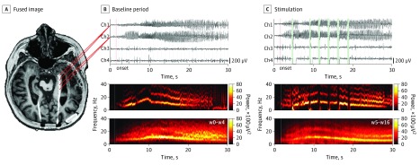
Figure 1. Closed-Loop Responsive Neurostimulation (RNS) Implantation and Data Processing Method.
A, Preoperative magnetic resonance image and postimplantation computed tomography fused image (EpiNav software27) aligned in the axial plane across the trajectory of the implanted RNS lead in the left hippocampus of patient 6. B, The 2 distal anterior hippocampal contacts provide bipolar channel 1, and the 2 proximal posterior hippocampal contacts form bipolar channel 2 (top), which record a unilateral electrographic seizure pattern (ESP) in the left hippocampus during the baseline period. For channel 2, a time-frequency plot shows the spectral evolution of the ESP aligned to onset (red vertical line). C, During stimulation, the amplifier is disconnected (time intervals in green), thereby generating a rectangular pulse artifact in the time domain (top) that is often followed by a considerable amplifier saturation direct current shift. In the frequency domain, stimulation appears as a low-frequency artifact accompanied by broadband cancellation (middle) that is often followed by a wideband artifact corresponding to amplifier saturation. Ch indicates channel; w, week of stimulation.

