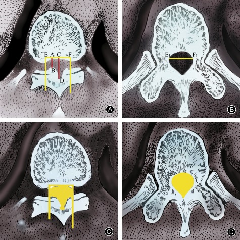
Figure 6.

Measurement of the residual spinal canal area based on CT scans. (A) AB and CD are the sagittal diameter and developing sagittal diameter,respectively, of the ossification segment. Spinal canal transverse diameter is calculated at the maximally stenosed level (E1 to F1). A vertical line extending through the endpoints of the transverse diameter (E and F) determines the boundary of the spinal canal. (B) The widest distance between two pedicles as viewed on a CT scan image of a transverse section through the pedicle of the same vertebra is measured as the spinal canal transverse diameter (E1 to F1). (C) Photoshop is used to measure the spinal canal area of the stenosed level. (D) A normal spinal canal area is measured by using the transverse section through the pedicle of the same vertebra.
