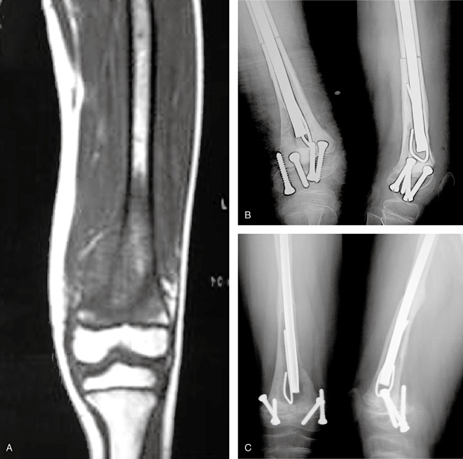
Figure 3.

A 6 year‐old girl with an osteosarcoma in the left distal femur. (A) An MRI image showing no involvement of the epiphysis. (B) An X‐ray image one week after surgery showing internal fixation in good position. (C) An X‐ray image 40 months after surgery showing bone healing with internal fixation in good position.
