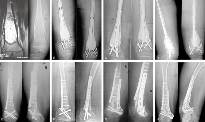
Figure 4.

An 8‐year‐old girl with an osteosarcoma in the right distal femur (A) MRI and X‐ray images show no involvement of the epiphysis (Type I). (B) Four days after surgery, the X‐ray image shows a visible gap between the host and inactivated bone with internal fixation in good position. (C) Three months after surgery, the gap has decreased with internal fixation in good position. (D) Two years after surgery, the inactivated bone was found to have fragmented with internal fixation still in good position. (E) One week after reoperation and implantation of allograft showing internal fixation in good position. (F) Twenty months after reoperation, the allograft has fragmented. (G) Three months after the third operation, the internal fixation and autograft are in good position. (H) Twenty‐eight months after the third operation, bone healing is visible in the X‐ray images with internal fixation in good position.
