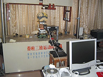
Figure 2.

Spine 2000, made in the Southern Medical University of China, including computer driven scan and mounting plates. These allowed for pure moment application of sagittal and axial rotation. The scan was mounted anteriorly across the L3‐L4 and L4‐L5 intervertebral discs and measured sagittal and axial rotation. Six small tridimensional iron boxes were used in the transverse processes as marked for scanning in lateral bending and torsion, and three in flexion‐extension.
