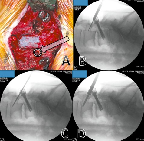
Figure 1.

L3–4 instrumented posterolateral fusion—previous L4‐S1 onlay fusion performed 12 years ago. (A) The starting point (arrow) is at the pars interarticularis, inferior to the mobile cranial facet joint. (B) Image intensifier image showing it use for directing the drill in the planned trajectory. (C) Image intensifier image showing that, because most of the of bone traversed in this technique is cortical bone, the hole is tapped up to the planned screw diameter. (D) Image intensifier image showing insertion of screw.
