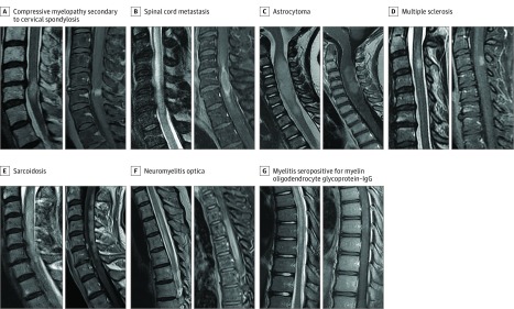
Figure 2. Gadolinium Enhancement Patterns in a Control Group With Alternative Causes of Myelopathy.
Control group of patients with alternative contrast-enhancing spinal cord lesions, with T2-weighted imaging on the left and accompanying T1-weighted imaging with gadolinium administration on the right. A, Compressive myelopathy secondary to cervical spondylosis, with a pancakelike band of enhancement just below the maximal area of stenosis. B, Spinal cord metastasis from squamous cell lung cancer with rim and flame enhancement. C, Astrocytoma demonstrating T2-weighted hyperintense mass with nonspecific accompanying enhancement. D, Multiple sclerosis with well-defined homogenous ovoid enhancing lesion in the dorsal spinal cord. E, Sarcoidosis displaying long dorsal subpial enhancement. F, Neuromyelitis optica with aquaporin 4–IgG seropositivity with a long contrast-enhancing spinal cord lesion and a component of central sparing, forming a ringlike enhancement. G, Myelitis with myelin oligodendrocyte glycoprotein–IgG seropositivity with a long T2-hyperintense lesion and faint patchy gadolinium enhancement.

