
Figure 1.
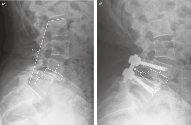
(A) Preoperative and (B) postoperative lateral radiographs demonstrating the various measurements used. (A) Preoperative measurements as follows: (a) L2‐S1 lordosis; (b) amount of anterolisthesis; and (c) cranial vertebral body width. (B) Postoperative measurements as follows: (a) posterior disc height; (b) distance of cage from anterior aspect of vertebral body; and (c) cranial vertebral body width.