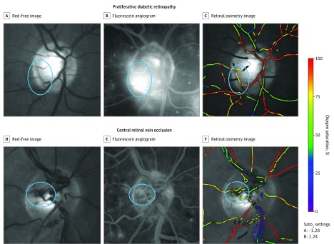
Figure. Oximetry of Disc New Vessels in Proliferative Diabetic Retinopathy and Disc Collateral Vessels in Central Retinal Vein Occlusion.
Red free (A), fluorescein angiography showing active leakage (B), and retinal oximetry imaging (C) of an eye with active proliferative diabetic retinopathy with disc new vessels. Red free (D), fluorescein angiography showing no leakage (E), and retinal oximetry imaging of an eye with central retinal vein occlusion and disc collateral vessels. The area of new vessels or disc collateral vessels is circled in all images. The panel on the right is the oxygen-saturation color bar. Sato2 indicates oxygen saturation.

