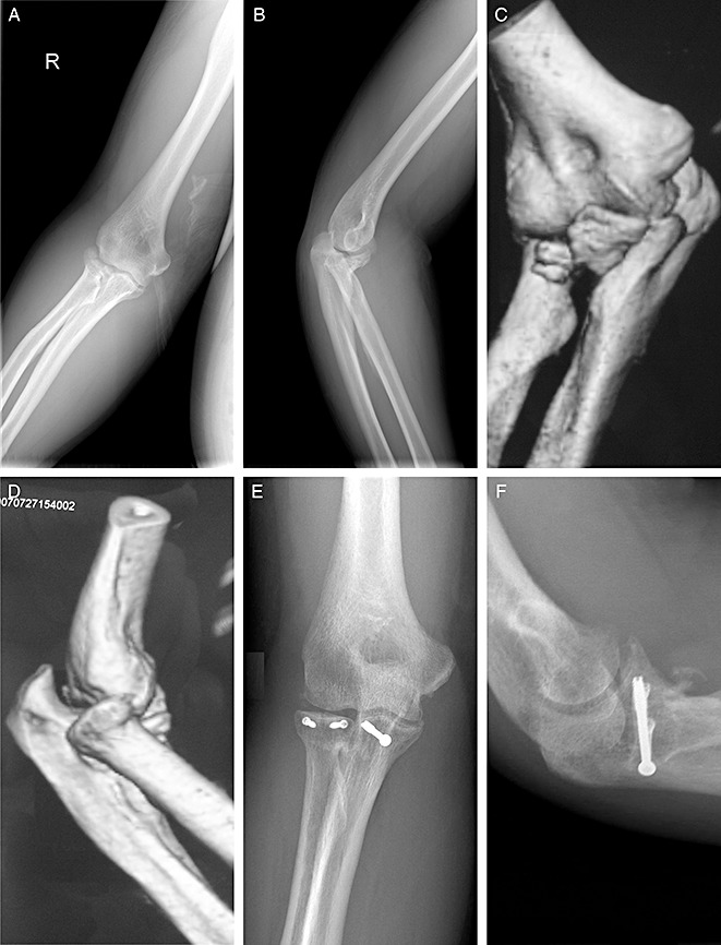
Figure 1.

A 52‐year‐old male patient with terrible triad of the elbow. (A, B) AP and lateral radiographs and (C, D) 3D CT reconstruction images reveal a radial head fracture (Mason type II), ulnar coronoid process fracture (Regan‐Morrey type II) and posterior dislocation of the elbow joint, and a small fragment of bone adjacent to the trochlea of the distal part of the humerus is a radial head fracture fragment. (E, F) AP and lateral radiographs taken 30 months postoperatively reveal achievement of a reduced and stable elbow joint. The radial head fracture was fixed with two Herbert screws, whereas the ulnar coronoid process fracture was fixed with a hollow nail. Slight heterotopic ossification and mild degenerative joint changes can be seen.
