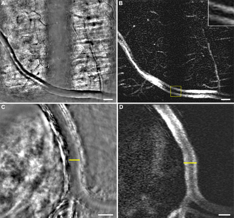
Fig. 3.
(A,C) Average images including vessels with a contrast typical of dark-field modality and (B,D) their respective perfusion maps (see Visualization 4 (24.2MB, avi) and Visualization 5 (9.1MB, avi) in Supplementary data). (A) Large vessel of peripheral retina lying on the nerve fiber layer alternatively illuminated in bright-field and dark-field configuration. A magnified view (yellow box) in (B) shows the aspect of the perfusion map on a large vessel when illuminated in dark-field. The large vessel shows two parallel bands, with a darker aspect in the center of the vessel, instead of the typical several bands observed in bright-field configuration (cf. Fig 2(D)). Again, capillaries hardly visible in (A) appear contrasted in (B). (C) Artery from the optic nerve head region. The artery is lying over the lamina cribrosa which illuminates it from behind leading to forward scattering which gives this contrast to the vessel, characteristic of dark-field illumination. In (D) the large vessel lumen is easier to measure than in (C) where it is hard to determine the beginning of the wall (yellow lines). Scale bars are 50 μm

