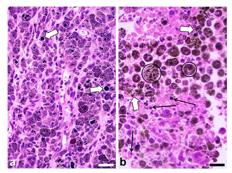
Fig. 4.
Histological H&E images of the melanotic melanoma B16-F10 growing in BALB/c mice, (a) without irradiation (control tumor), and (b) after irradiation with the 808-nm laser for 10 min and observed 24 h later. (a) Small and large black granules (melanosomes) appear within tumor cells, but sometimes they are located in the extracellular space (white arrows). The irradiated tumor section (b) shows extensive damaged and necrotic areas with rounded or disrupted tumor cells (white arrows), big brown-black melanophages (encircled), as well as pycnotic cell nuclei (black arrows), and cytoplasm fragmentation. Scale bars: 30 µm.

