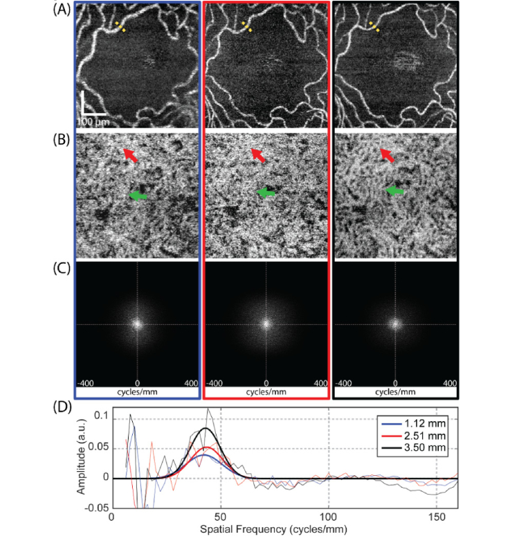
Fig. 4.
The clarity of the OCTA vascular image is affected by the incident beam size. (A) En-face retina OCTA images at FAZ region imaged by the beam diameters of 1.12 mm, 2.51 mm and 3.50 mm (from left to right), respectively. (B) Corresponding en-face CC OCTA images. (C) 2D power spectrums of en-face CC OCTA images for all three beam diameters. (D) Offset-removed radially-averaged power spectrums of CC (solid line, thin, blue for the beam diameter of 1.12 mm, red for the beam diameter of 2.51 mm and black for the beam diameter of 3.50 mm) and their corresponding Gaussian fitting curves (solid line, bold).

