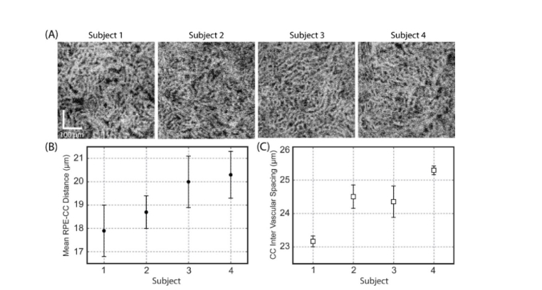
Fig. 5.
The choriocapillaris lobular network under the central fovea region can be resolved by a commercial grade SS-OCT system with an incident probe beam diameter of ~3.50 mm at the pupil plane. (A) En-face OCTA images of the CC slab at the posterior pole region acquired from 4 normal subjects. (B) Measured mean RPE-CC distance (mean ± std) of the 4 subjects. (C) Measured mean CC inter-vascular spacing (mean ± std) of the subjects.

