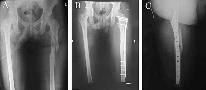
Figure 5.

Radiographs of a patient (Group B) with a comminuted subtrochanteric fracture treated by the reverse Liss‐DF technique; (A) Before surgery; (B) One week after surgery: satisfactory fracture reduction and stable fixation can be seen in the AP and (C) lateral views.
