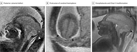
Figure 3. Fetal Magnetic Resonance Imaging Performed at 28 and 37 Weeks’ Gestational Age.
A, Sagittal image at 28 weeks’ gestational age shows a broad posterior calvarial defect through which the posterior cerebral hemisphere herniates (black arrowhead), consistent with an encephalocele. Extracerebral and posterior fossa subarachnoid fluid spaces are absent; the fourth ventricle is not visualized, typical of stigmata of a Chiari II malformation (white arrowhead). Severe microcephaly is evident. B, Axial image at 28 weeks’ gestational age near the vertex shows the posterior protrusion of cerebral hemisphere through the calvarial defect (arrowheads) and lack of cerebrospinal fluid around the cerebral hemispheres. C, Sagittal midline image at 37 weeks’ gestational age demonstrates similar findings of a posterior encephalocele (arrowheads) and stigmata of a Chiari II malformation as evident in panel A.

