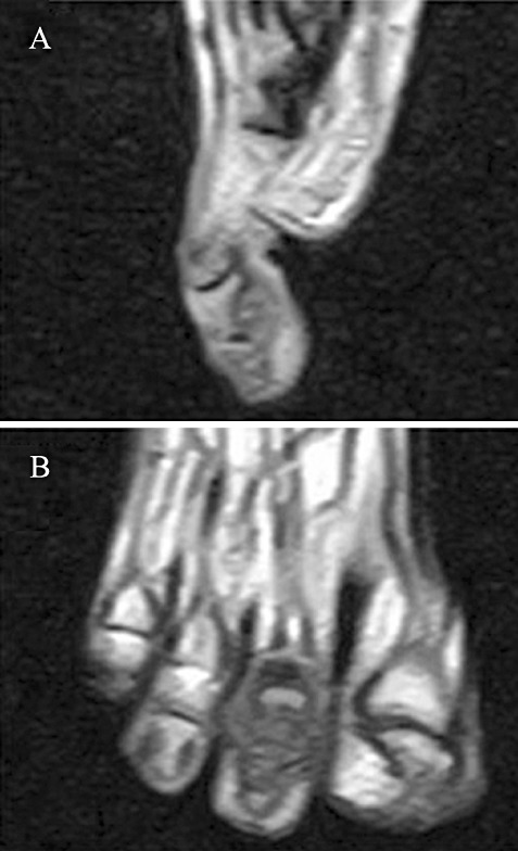
Figure 8.

(A) Sagittal plane MRI displaying a soft‐tissue tumor on the plantar aspect of the second toe with a characteristic low signal on T1weighted image. (B) Coronal plane MRI showing the soft‐tissue tumor separated by a pseudomembrane and bulging on both sides of the second toe.
