
Figure 4.
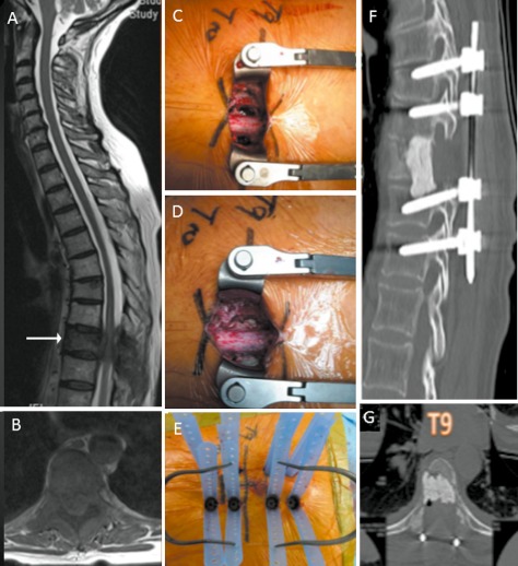
A 58‐year‐old woman with lung cancer presented with midthoracic pain of 2 months duration. (A) and (B) MRI images showing circumferential spinal cord compression at T9 level. (C) Intraoperative photograph showing mini‐open midline linear incision with bilateral pediculectomy and vertebrectomy. (D) Post vertebroplasty with cement augmentation. (E) Intraoperative photograph showing percutaneous sleeves in situ. (F) and (G) Post‐operative CT scan images.